Diagnostic imaging
Our hospital boasts a vast array of diagnostic imaging equipment. These include endoscopy, radiography, ultrasonography, nuclear scintigraphy (‘bone scan’), and magnetic resonance imaging (MRI).
About Diagnostic imaging
Advances in diagnostic imaging have revolutionised our ability to accurately diagnose, treat and monitor injury and disease in horses. Diagnostic imaging allows us to visualise structures, and activities, within the body.
At Three Counties Equine Hospital we have a vast array of both portable and hospital based diagnostic imaging equipment. These include:
Radiography
Ultrasonography
Nuclear scintigraphy (‘Bone Scan’)
Magnetic resonance imaging (MRI).
Many problems are effectively investigated and monitored at the horse’s own yard by our team of vets, using portable digital radiography and ultrasonography equipment. However, for more in-depth and complicated investigations, horses and ponies can be brought into the hospital to utilise the more advanced imaging equipment.
Each imaging modality provides evaluation of different structures and may be used alone, or in combination, to achieve a diagnosis. Our team of specialised and experienced vets will discuss these at the time and select the most appropriate and suitable imaging technique to increase the likelihood of achieving a diagnosis.
Radiography
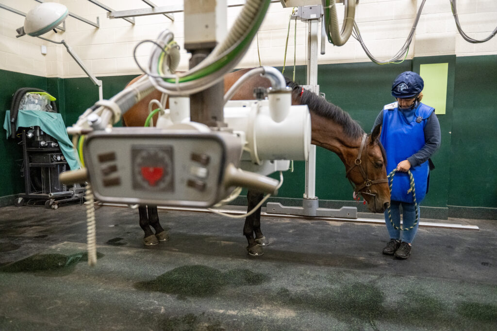
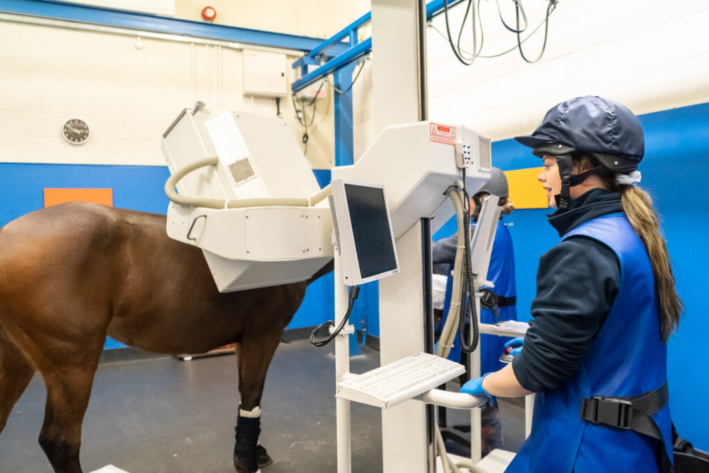
We use a hospital based high powdered X-ray generator which can obtain high quality images of the limbs, head, neck, back, chest and abdomen.
We also have several mobile systems that can be transported to the yard for imaging out and about as required.
These days radiography is all digital, whereby the x-ray beam is converted to an electronic image almost instantly and can be manipulated to provide the best quality images. These can then be easily shared with owners, farrier sea other vets as required.
Ultrasonography
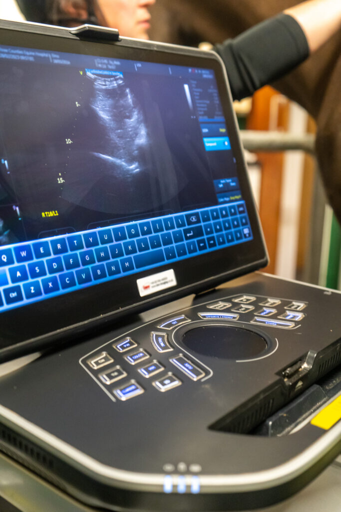
Ultrasound is a very useful imaging technique that uses real time sound waves to image different parts of the body. We have many ultrasound machines used for different imaging modalities such as orthopaedic imaging of limbs, neck and back, abdominal and thoracic imaging of medical cases and colics, heart scanning for investigation of murmurs and arrhythmias, reproductive imaging for breeding mares and detecting and monitoring pregnancies. Other parts of the body such as eyes, masses, foreign bodies are routinely imaged with ultrasound.
Some machines are large and hospital based and others are mobile and used on the road.
Nuclear scintigraphy
Nuclear scintigraphy (bone scanning) involves injection of a radioactive marker which accumulates in bone and soft tissue identifying sites of injury. This can sometimes show up bone injury before it is evident on radiographs. Bone scans are performed with the horse standing and sedated, and take around 2-3 hours depending on how much of the horse is being scanned.
Bone scans are very useful for detecting stress fractures, for multi-limb lameness cases, for horses who are difficult to nerve block, or where injury is suspected in areas that are hard to image otherwise, such as the pelvis and back.
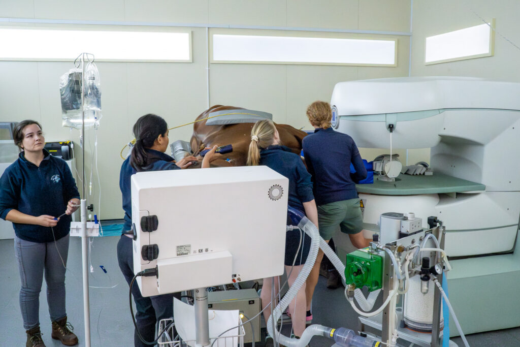
MRI
Our MRI is an open magnet with a large field of view. In addition to routine MRI of the lower limbs (feet, pasterns, fetlocks, suspensories) it is the only machine in the country capable of imaging difficult-to-reach areas, such as the upper limbs (knees, hocks, stifles) and the head.
As our MRIs are performed under general anaesthesia, we can obtain superior imaging quality by removing any artefact caused by the horse moving.
We are here to help
Reach out to our team with any questions or to arrange an appointment — we are always happy to hear from you.