
Magnetic resonance imaging / ‘MRI’
MRI is a useful technique for diagnosing and evaluating soft tissue and bone injuries. At TCEH we have a large open MRI scanner. It is the first of its kind in the country, and the only one in the UK capable of scanning stifles, as well as accurately scanning heads, knees, suspensories, hocks, fetlocks and feet.
The MRI is carried out under general anaesthetic which eliminates any movement in the region being scanned and therefore produces superb image quality. The high quality of the images produced means we can diagnose pathology in structures early on in the course of disease/injury therefore allowing us to tailor more specific treatment and rehabilitation programs, hopefully producing better outcomes for the patient. The scans also help us to know when treatment attempts may be futile, saving time and unnecessary disappointment.
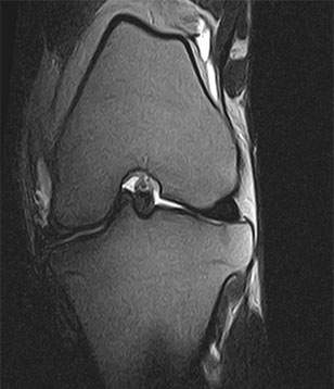
Dorsal stifle
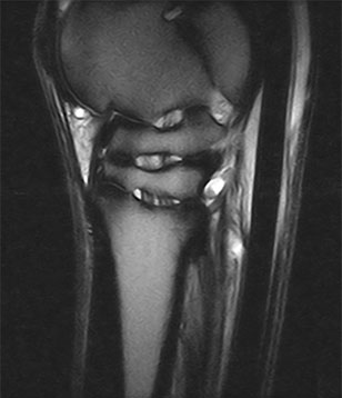
Saggital hock/Suspensory
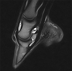
Saggital foot
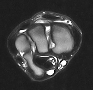
Transverse carpus
Nuclear scintigraphy / ‘Bone Scanning’


Scintigraphy involves injecting the horse with a radioactive marker that is taken up by the bone and highlights areas of abnormal boney activity. This is very useful in evaluating conditions, which are difficult to identify using other imaging techniques. It proves very useful in assessing problems of the neck, back and pelvis, for evaluating multi-limb lameness, for diagnosing stress fractures in racehorses, and for assessing horses which are difficult to nerve block.
Radiography
Radiography at the hospital is performed using our powerful ceiling-mounted X-ray machine that generates high quality, digital images. Although its main use is for lameness and back pain investigation, it also plays an important role in the evaluation of dental pathology, head and neck problems and thoracic disease. We also have a mobile digital radiography system for use in the field.
Ultrasonography
The practice owns several digital diagnostic ultrasound scanners, all of which are portable.
Ultrasound is used routinely for examination of the reproductive tract in the breeding mare. It is also used widely for orthopaedic problems to evaluate tendon, ligament and joint problems. It is also invaluable when assessing neck, back and sacroiliac problems, and to guide injections at these sites.
Ultrasonography is also used for evaluating the thorax and abdomen, such as assessing colics, liver disease and respiratory problems. It is also used for heart scanning (echocardiography).
Videoendoscopy and videogastroscopy
Our various endoscopes allow direct visualisation of patients’ internal organs such as the upper and lower airway, sinuses, oesophagus, stomach, duodenum and bladder.
With videoendoscopy, highly detailed images are projected onto a television monitor, which allows clients to see the images themselves and gives improved understanding of the problem and proposed treatment options.
Laparoscopic Surgery
Laparoscopy is an advanced surgical technique which allows certain procedures in the horse to be performed minimally invasively (through small incisions). Many of these procedures can be performed in the standing horse, negating the need for a general anaesthetic.
There is a vast range of surgeries that can be performed laparoscopically including chryptorchidectomy (‘rig’) surgeries and ovariectomy.
Ovariectomy
The most common indication for ovariectomy in the horse is development of a tumour within one of the ovaries – most commonly a ‘granulosa cell tumour’. These are benign tumours but may grow to a large size that may interfere with other organs and affect ridden performance. They secrete a large array of hormones which can cause stallion like behavior or the mare not to cycle. Diagnosis is commonly made on a combination of ultrasound examination of the ovary and hormone analysis. The surgery is most commonly performed in the standing sedated patient using local anaesthetic.
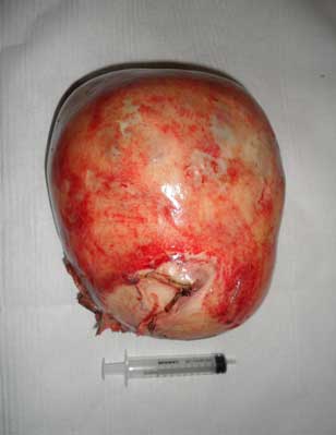
A large pathological ovary following removal

An abdominally retained (left) and normally descended testes following removal
Cryptochidectomy
A cryptorchid or ‘rig’ is where one or both of the testes are retained within the abdominal cavity. Commonly these horses will show stallion like behavior that is often undesirable. It is possible to identify the position of the testis using ultrasonography which can assist with the procedure. Testes that are abdominally retained are better candidates for laparoscopy than those with inguinally retained testes.
Similarly to ovariectomy the procedure is most commonly performed in the standing sedated patient, but can also be performed under general anaesthetic.